Microscopy
The microscopic nature of matter is investigated at DiSC by a wide type of methods, ranging from the Scanning Probe Microscopy (SPM) to the Optical Microscopy.
SPM covers several related techniques that, using a probe that scans the specimen, image surfaces and measure surface properties on a fine scale (down to the molecular/atomic scale). SPM techniques include Scanning Tunneling Microscopy (STM), Atomic Force Microscopy (AFM) and Magnetic Force Microscopy (MFM).
In particular here can be reported:
NT-MDT SPM (AFM, MFM, STM ...) solver P47H-PRO
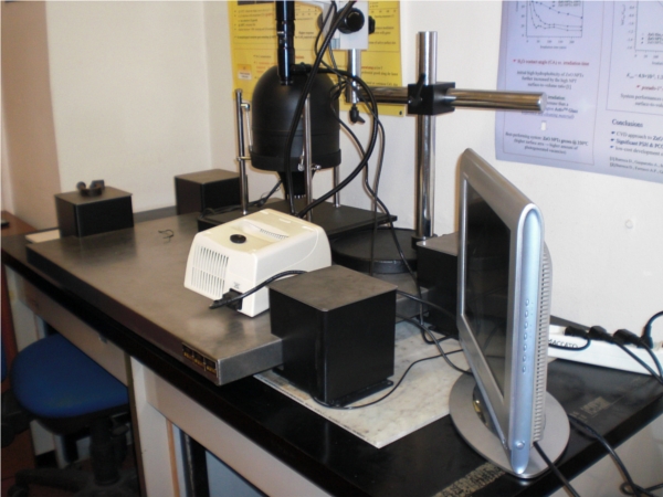
This AFM allows for the production of high quality images from a wide variety of materials. Typically, the spatial resolution achieved ranges in the order of fractions of a nanometer, thus enabling for the visualization of atomic and molecular packings. The equipment includes a complete set of tips, enabling the contact, non contact and tapping operation modes. In the tapping mode the intermittent contact frequency can be set by the user. Conductivity maps or surface potential images can be produced from electrically conducting materials. Magnetostatic interactions between suitable tips and samples hosting magnetic domains can be studied, with a resolution up to 10 nm. The AFM is interfaced to an optical microscopy equipped with a CCD camera, to monitor the laser alignment on the tip and the sample surface. This AFM is actually applied to the realization of detailed snapshots of samples kept in air, without any need of high vacuum conditions or sputtering pre-treatments. This allows for the study of degradable and soft materials.
contacts: chiara.maccato@unipd.it
location at DiSC: 215-00-044
Agilent 5500 AFM/STM
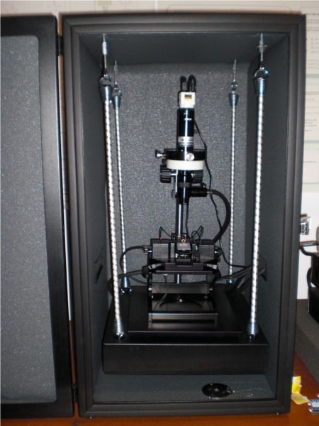
The Agilent 5500 Atomic Force Microscope/Scanning Tunnelling Microscope (AFM/STM) system housed at DiSC can be operated in contact, non-contact and tapping modes. Beyond the more traditional surface studies, liquid samples can be analysed. Conductivity maps of conducting materials can be determined. The instrument can provide phase-contrast imaging, bearing additional and complementary information with respect to the more conventional topographic contrast set-up. In the STM mode the instrument affords the control of current set-point levels as low as few pico-Amperes. This AFM/STM is equipped with an eight-gate environmental chamber that allows to maintain the sample and the tip under controlled atmosphere during measurement, thus enabling dynamic studies characterized by controlled changes of the gas or liquid ambient surrounding the sample in the scan-time, with no need to stop the run.
contacts: mauro.sambi@unipd.it
location at DiSC: 215-02-083
Zeiss SUPRA 35VP Scanning Transmission Electron Microscopy (STEM) equipped with an Oxford INCA X-Sight detector
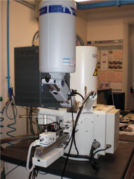
This STEM is characterized by the high level of the electronic beam collimation, guaranteed by the In-lens systems in column, which allow for the in-column collection of the signals from back-scattered and secondary scattered electrons, thus avoiding any parallax error and allowing for the production of images approaching the few nanometers spatial resolution scale, in the best conditions.
The system is equipped with a Field Emission cathode, a GEMINI electron-optics column and four detectors: 1) In-Lens secondary electrons detector; 2) Everhart-Thornley secondary electrons detector; 3) Variable Pressure secondary electrons detector (VPSE); 4) four quadrants backscattered electrons detector (AsB) EDX for elemental analyses, interfaced to a Oxford Instruments Inca X-Act EDX spectrometer. The system allows for oil-free high vacuum (5x10⁻⁷ torr) operating mode and variable pressure condition, from 1Pa to 144 Pa, thus allowing for investigations on wet samples.
The SmartSEM software allows for detailed analyses of the experimental data.
contacts: chiara.maccato@unipd.it
location at DiSC: 215-00-132
LEICA DM4000M optical microscopy
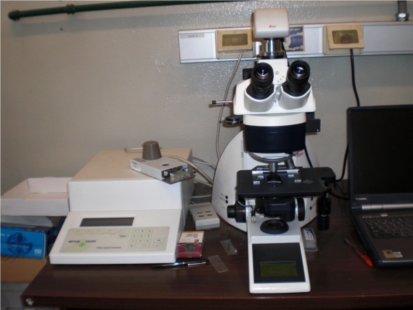
The Leica DM4000M optical microscope housed at DiSC is equipped with light polarizers, a photocamera interfaced to a computer by a versatile acquisition software for fast collection and automatic archiving of images and a Mettler Toledo FP82HT Hot Stage for the temperature control of the samples. Materials can be supported on a conventional microscope slide and heated up to 250°C or thermostated to a set temperature with the precision of ± 0.1°C. Details on the tens of micrometers length scale can be detected by the 50x magnification optics. The system is particularly suitable for the study of polymorphism features, as well as morphology and kinetics of phase transitions in complex polymeric materials. The microscope is actually dedicated to materials science and forensic research, in particular polymeric film and fiber characterization, inks and documents dating.
contacts: carla.marega@unipd.it
location at DiSC: 215-01-073
Axiotech Zeiss optical microscope
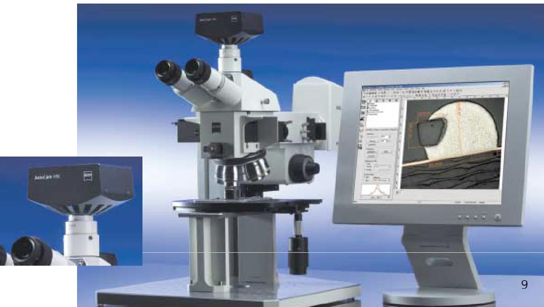
An Axiotech Zeiss optical microscope equipment provided with lenses up to 500 magnifications and dichromator with registration camera is actually applied for the investigation of research samples and for Cultural Heritage Artefacts. A microscope that combines precision, stability, and flexibility. Its name: Axiotech, works also with non-standard size samples. It features a large continuously adjustable specimen area and baseplate with a grid pattern of holes –guaranteeing you maximum flexibility for needs and experimental set-ups. It has the option of precisely determining sizes laterally and vertically with the 3-axis measuring system from Carl Zeiss. In addition it is integrated in a digital imaging platform.
contacts: renzo.bertoncello@unipd.it
location at DiSC: 215-03-048
Olympus CX 41 and Olympus SZ30 optical microscopes
 Routine sample observations can be performed by some Olympus optical microscopes housed at DiSC.
contacts: tommaso.carofiglio@unipd.it ; renato.schiesari@unipd.it
location at DiSC: 170-00-010 ; 215-00-132
note: fluorescence microscopy techniques are listed under "Optical Spectroscopy"